
Facts & figures about skin cancer


The next graphic is an illustration of our skin. To the very left there is a healthy mole. When a healthy mole transform to a cancerous melanoma, it first increases its size in the epidermis. In that stage, the cancer can be removed quite easy so that no cancer cells remain in your body.
When the melanoma keeps growing, it goes deeper into the dermis. Its getting harder to remove the cancer entirely, so an additional radiation and/or chemotherapy might be necessary.
When the cancer grows even further and reaches the bloodstream, cancer cells can easily spread over your hole body and get into your lymph system. It will start to grow metastasis in your bodies organs. The chances to cure skin cancer with metastasis are very low.

The solution - PHILIPS SkinScan
To allow a 3D scan of your body, there is a laser, a camera and a laser receiving sensor necessary, like already used in todays 3D scanners.
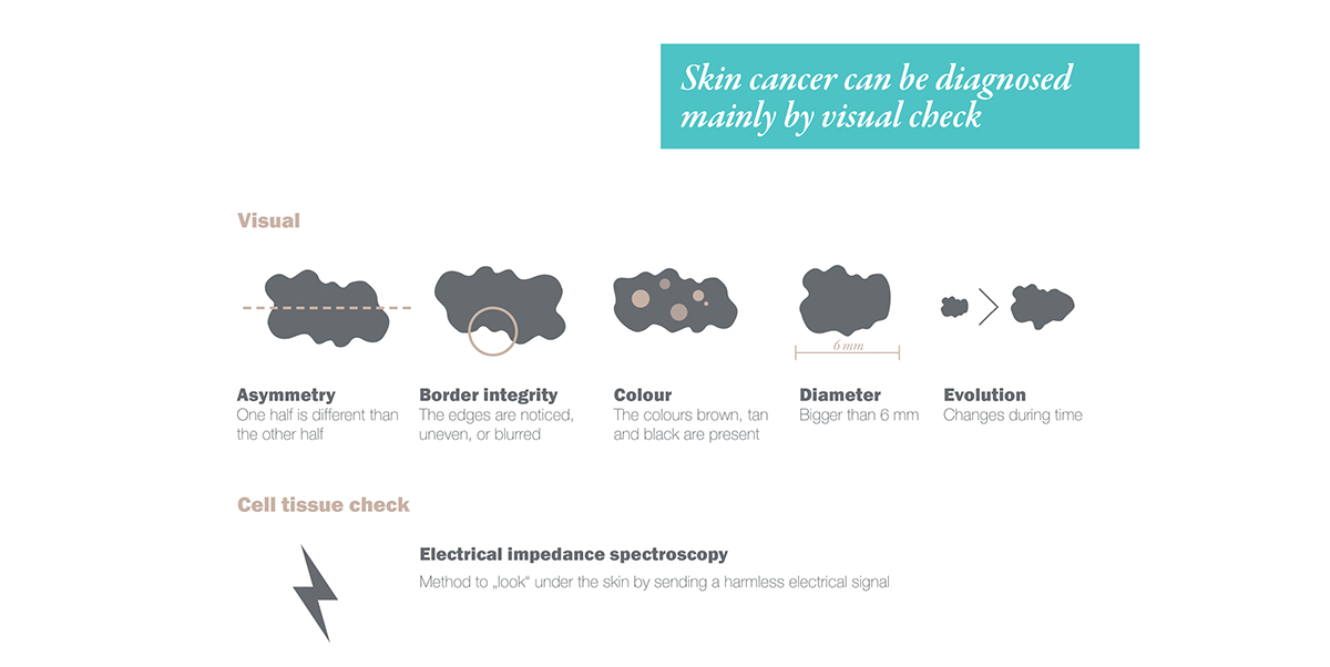
These components are covered and protected by a glass. Embedded in this glas, there are electrodes, that can send an electric current to your skin, that is analysed, to double check on the skin cells set up.
The glass is surrounded by an LED ring. The light improves the quality of the captured images. An exact colour analysis is necessary to detect cancer.


Usage scenario


For more information check out the full project report:


